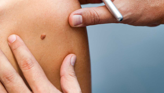
Basal cell carcinoma (BCC) is the most common type of skin cancer. This malignant tumor is locally invasive, aggressive, and destructive, but there is a limited capacity to metastasize. The reason for this characteristic is the tumor’s growth dependency on its stroma, which on invasion of tumor cells into the vessels is not disseminated with the tumor cells. When tumor cells lodge at distant sites, they do not multiply and grow because of the absence of growth factors derived from the stroma of the tumor. Exceptions occur when a BCC shows signs of dedifferentiation, for instance, after inadequate radiotherapy. BCC usually arises only from epidermis that has a capacity to develop (hair) follicles. Therefore, BCCs rarely occur on the vermilion border of the lips or on the genital mucous membranes.
Most lesions are readily controlled by various surgical techniques or cryotherapy. Serious problems, however, may occur with BCC arising in certain locations on the face: around the eyes, in the nasolabial folds, around the ear canal, or in the posterior auricular sulcus. In these sites the tumor may invade deeply, cause extensive destruction of muscle and bone, and even invade to the dura mater. In such cases, death may result from hemorrhage of eroded large vessels or infection (meningitis).
Causes of Basal Cell Carcinoma
- The sun is responsible for over 90 percent of all skin cancers, including BCC, and chronic overexposure to the sun is the cause for most cases of basal cell carcinoma. BCCs – the tumors themselves – occur most frequently on the face, ears, neck, scalp, shoulders, and back.
- UV radiation: This is the most important and common cause of BCC.
- Other risks include older age, genetic predisposition (skin cancers are more common in those who have light-colored skin, blue or green eyes, and blond or red hair), chemical pollution, and overexposure to x-rays or other forms of radiation.
Symptoms of Basal Cell Carcinoma
- A skin lesion, growth, or bump located on the face, ear, neck, chest, back, or scalp and it appear like Pearly or waxy appearance, White or light pink, flesh-colored, or brown, Flat or slightly raised.
- Visible blood vessels in the lesion or adjacent skin
- Appearance of a scarlike lesion without a history of injury to the skin in that area
- A sore that will not hea
- Surface may be scaly or crusted
Diagnosis
Serious BCCs occurring in the danger sites [central part of the face, behind the ears] are readily detectable by careful examination with good lighting, a hand lens, and careful palpation.
Treatment
Excision with primary closure, skin flaps, or grafts. Cryosurgery and electrosurgery are options, but not in the danger sites or on the scalp.
For lesions in the danger sites (nasolabial area, around the eyes, in the ear canal, in the posterior auricular sulcus, in sclerosing BCC, and on the scalp) microscopically controlled surgery (Mohs surgery) is the best approach. Radiation therapy is an alternative only when disfigurement may be a problem with surgical excision (e.g., eyelids or large lesions in the nasolabial area).
There are a variety of topical treatments that can be used for superficial basal cell carcinomas but only for those tumors below the neck; this is because these therapies are not always definitive in removing all the neoplastic cells, and recurrences on the head and neck may be difficult to manage because of the invasion along the fascial planes. Nevertheless, these modalities do save the need for surgery with its resultant scarring and are worthwhile but only when used on the arms, legs, and trunk. Some patients prefer cryosurgery, but this treatment leaves a white spot that remains for life. Electrocautery and curettage is another popular treatment, but it leaves scars. Topical 5-fluorouracil ointment and imiquimod cream are effective and do not cause scars. Some new modalities are promising, such as PDT (photodynamic dye + visible light), and this does not leave a scar.
References