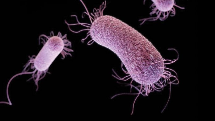
Pseudomonas aeruginosa exists in moist environments associated with hospitals. Hospitalized compromised individuals become colonized with the organism. Local invasion can follow colonization of any mucocutaneous site with local infection and/or hematogenous dissemination. Ecthyma gangrenosum (EG) is the necrotizing soft-tissue infection that occurs after local tissue invasion or bacteremic seeding, associated with blood vessel invasion, septic vasculitis, vascular occlusion, and infarction of tissue.
Causes of Cutaneous Pseudomonas Aeruginosa Infections
P.aeruginosa rarely causes disease in the healthy host. Infections occur when normal cutaneous or mucosal barriers have been breached or bypassed (e.g., burn injury, penetrating trauma, surgery, endotracheal intubation, urinary bladder catheterization, IV drug abuse); when immunologic defense mechanisms compromised (e.g., by chemotherapy-induced neutropenia, hypogammaglobulinemia, extremes of age, diabetes mellitus, cystic fibrosis, cancer, HIV disease); when protective function of normal bacterial flora has been disrupted by broad-spectrum antimicrobial therapy; and/or when patient has been exposed to reservoirs associated with hospital environment. Infections caused by P.aeruginosa begin with superficial colonization of cutaneous or mucosal surfaces and progress to localized bacterial invasion and damage of underlying tissue. Infection may remain anatomically localized or may spread by direct extension to contiguous structures. Blood vessel and bloodstream invasion, dissemination, systemic inflammatory-response syndrome (SIRS) (sepsis syndrome), multiple-organ dysfunction, and, ultimately, death may follow localized infection. The organism and/or its products may cause tissue injury at primary and secondary sites of infection; release of systemically acting toxins or inflammatory mediators of infected host may contribute directly or indirectly to SIRS.
Diagnosis
Clinical suspicion confirmed by blood and skin exudate/biopsy specimen culture.
Treatment
Correct Predisposing Factors White cell transfusion or G-CSF for granulocytopenia.
Antimicrobial Therapy Antibiotic and dosage are adjusted according to sensitivities and results of cultures.
Surgery After control of infection, areas of infarction should be debrided.
References