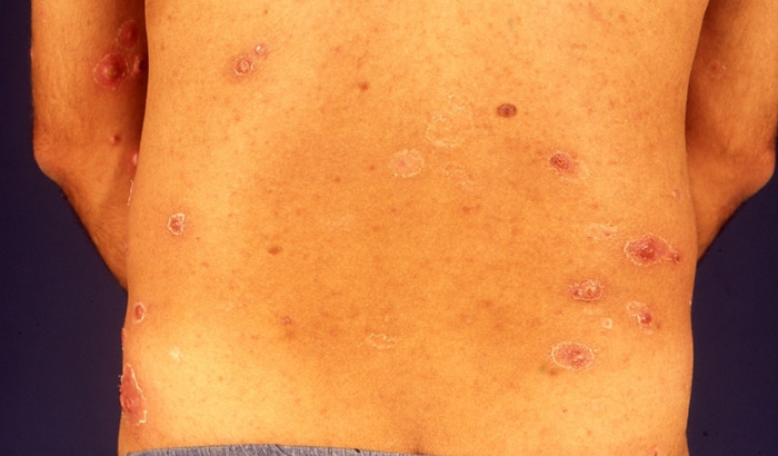
Lymphomatoid papulosis is a stubborn persistent rash that usually occurs on the chest, stomach, back, arms and legs.
Lymphomatoid papulosis is an asymptomatic, chronic, self-healing, polymorphous eruption characterized by recurrent crops of lesions that regress spontaneously, with histologic features of lymphocytic atypia. It is a low-grade, self-limited T cell lymphoma with a low but real risk of progression to more malignant forms of lymphoma.
Causes of Lymphomatoid Papulosis
Unknown; considered to be a low-grade lymphoma controlled by host mechanisms without systemic involvement. Antigens shared with and occasional progression to Hodgkin’s disease or cutaneous T cell lymphoma suggest a low-grade lymphoma, perhaps induced by chronic antigenic stimulation. Lymphomatoid papulosis may begin as a chronic, reactive, polyclonal lymphoproliferative phenomenon that sporadically overwhelms host immune defenses and evolves into a clonal, antigen-independent, true lymphoid malignancy. Belongs in the spectrum of primary cutaneous Ki-1 + lymphoproliferative disorders, including pseudo-Hodgkin’s disease of the skin, regressing atypical histiocytosis, Hodgkin’s lymphoma, and Ki-1 + large cell lymphoma.
Symptoms of Lymphomatoid Papulosis
Most patients present with multiple skin papules (raised bumps) that may occur anywhere on the body but most often affect the chest, stomach, back, arms and legs. The papules appear in crops and may be mildly itchy. They may develop into blood or pus-filled blisters that break and form a crusty sore before healing completely. Lesions spontaneously heal with or without scarring within 2-8 weeks of appearing.
Diagnosis
Based on typical histology and immunohistochemistry, lack of systemic involvement by history and physical examination. May necessitate periodic biopsy to rule out blastic transformation, especially with tumor formation. No reliable histologic, immunohistochemical, or genotypic test available for prediction of risk of progression to lymphoma.
Treatment
No treatments have proved consistently effective, as is evidenced by the multiple reported therapies. Topical agents include glucocorticoids and carmustine (BCNU). Electron-beam irradiation has been employed as well. PUVA may control the disease but does not affect the long-term prognosis. Tetracyclines, sulfones, systemic glucocorticoids, and even acyclovir have been reported as effective by some researchers. A wide spectrum of systemic agents has been used, including retinoids, methotrexate, chlorambucil, cyclophosphamide, cyclosporine, and interferon-α2b, none with lasting effect.
References