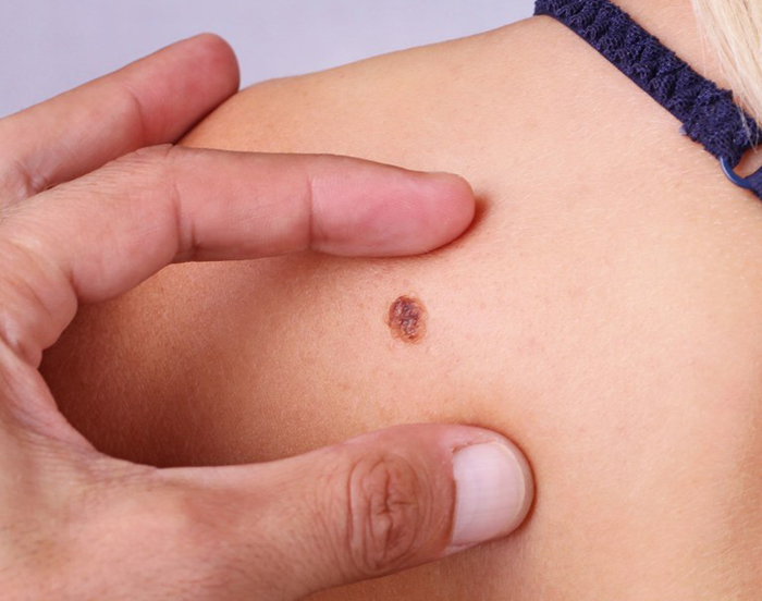
Merkel cell carcinoma (MCC) (cutaneous neuroendocrine tumor) is a rare malignant solid tumor thought to be derived from a specialized epithelial cell, the Merkel cell. It is a nondendritic, nonkeratizing, “clear” cell present in the basal cell layer of the epidermis, free in the dermis, and around hair follicles as the hair disk of Pinkus. The etiology is unknown, but may be related to chronic ultraviolet radiation (UVR) damage. The tumor may be solitary or multiple and occurs on the head and on the extremities. There is a high rate of recurrence following excision, but, more important, it spreads to the regional lymph nodes in more than 50% of the patients and can be disseminated to the viscera and CNS.
Causes of Merkel Cell Carcinoma
Ultraviolet radiation has been implicated as a factor in developing Merkel cell carcinoma, due to the occurrence of the tumour on sun exposed skin. Immunosuppression following organ transplantation is also associated with the development of Merkel cell carcinomas.
Symptoms of Merkel Cell Carcinoma
The first sign of Merkel cell carcinoma is a fast-growing, painless nodule (tumor) on your skin. The shiny nodule may be skin colored or may appear in shades of red, blue or purple. Nearly half of Merkel cell carcinomas appear on the face, head or neck, but they can develop anywhere on your body, even on areas not exposed to sunlight.
Diagnosis
- Physical exam. Examining unusual moles, freckles, pigmented spots and other growths on your skin is the first step your doctor will likely take in making a diagnosis.
- Biopsy. After removing the tumor or a sample of the tumor from your skin, your doctor treats the cells with a special stain for viewing under the microscope.
- X-ray
- Sentinel node biopsy
Treatment
Excision by Mohs’ surgery and prophylactic regional node dissection are advocated because of the high rate of regional metastases. In one series, even without a local recurrence, a large number of patients (60%) developed regional node metastases, and in those patients with local recurrence, 86% developed regional node metastases. Therefore, the excision should be followed by prophylactic radiotherapy to the nodal areas.
References